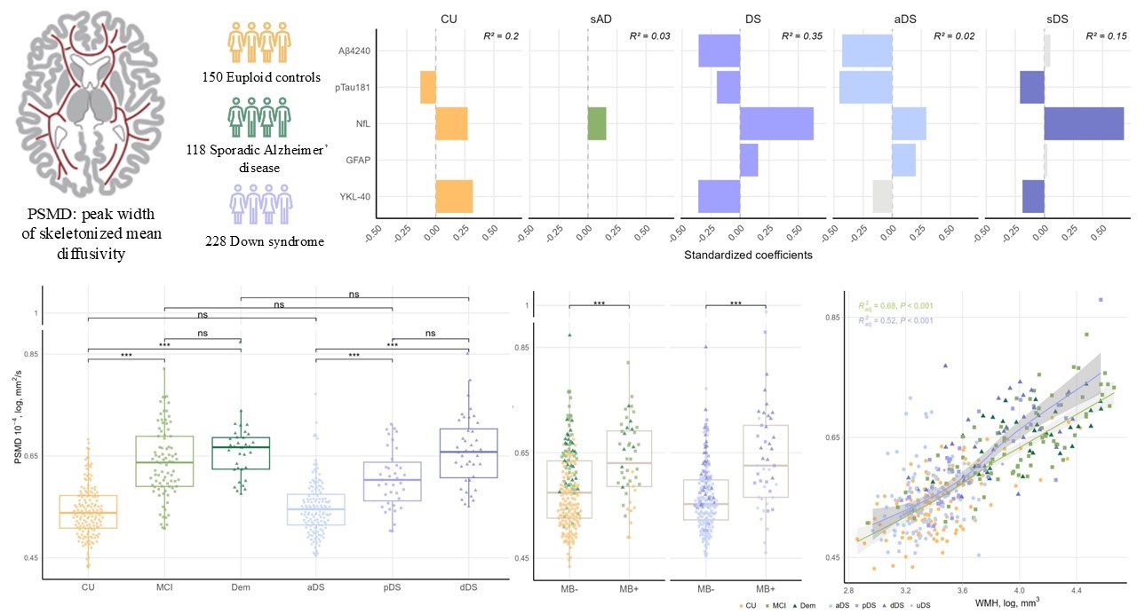
Peak width of skeletonized mean diffusivity reveals early and multifactorial white matter injury across sporadic and Down syndrome–associated Alzheimer’s disease

White matter alterations are commonly observed in Alzheimer’s disease (AD) and reflect a convergence of several pathological processes, including myelin loss, axonal degeneration, and microvascular injury. Peak width of skeletonized mean diffusivity (PSMD) is a diffusion MRI–derived metric of diffuse white matter injury that has demonstrated high sensitivity to microstructural damage. However, the timing of … Read more

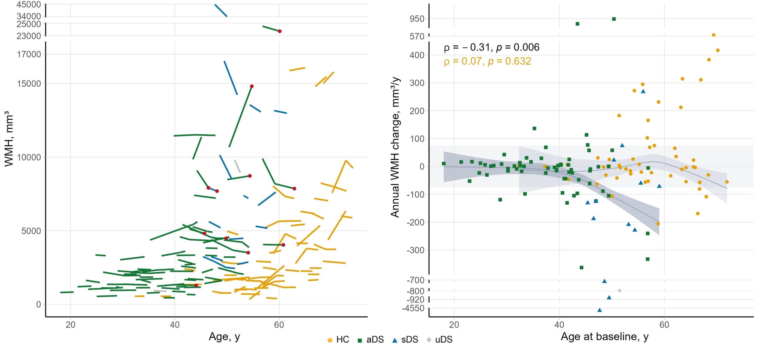
Temporal dynamics of white matter hyperintensities related to Alzheimer’s disease in adults with Down syndrome

Temporal dynamics white hyperintensities Down Syndrome - Sant Pau Memory Unit - Alzheimer - Barcelona

White matter hyperintensities (WMH) White matter hyperintensities (WMH) are brain lesions visible on MRI that have traditionally been linked to vascular disease. In a previous study from our group, we showed that WMH burden increases approximately 10 years before the onset of Alzheimer’s disease (AD) symptoms in people with Down syndrome (DS) and continues to … Read more

Study of brain perfusion in adults with Down syndrome along the Alzheimer’s disease continuum

Cerebral perfusion is the amount of blood reaching a given brain region at any given time. In other words, it is the “irrigation” that supplies neurons with oxygen and nutrients. When perfusion decreases or becomes abnormally distributed, it may indicate that an area is beginning to deteriorate, even before clinical symptoms or signs of atrophy … Read more

Brain Metabolism in Alzheimer’s Disease Associated with Down Syndrome: New Findings with FDG-PET

Regional Brain Metabolism across the Alzheimer's Disease Continuum in Down Syndrome

Down syndrome (DS) is a genetic form of Alzheimer’s disease (AD). Due to the overexpression of the APP gene on chromosome 21, nearly all individuals with DS develop AD-related pathological changes and dementia secondary to this disease. Brain metabolism, measured using fluorodeoxyglucose positron emission tomography ([¹⁸F]FDG-PET), is considered a biomarker of AD-related neurodegeneration. This recent … Read more

Medial Temporal Lobe Atrophy in Down Syndrome Along the Alzheimer’s Disease Continuum

Medial Temporal Lobe Atrophy in Down Syndrome Along the Alzheimer’s Disease Continuum The medial temporal lobe (MTL) is one of the first regions affected by tau pathology, making structural changes in these areas promising biomarkers for identifying early signs of Alzheimer’s disease (AD). However, little is known about the integrity of these regions in individuals … Read more

Enhanced Resolution to Study Synaptic Tau Pathology in Alzheimer’s Disease

Enhanced Resolution to Study Synaptic Tau Pathology in Alzheimer’s Disease

Our recent study shows that combining Array Tomography with an advanced imaging technique called Two-Colour Direct Stochastic Optical Reconstruction Microscopy (dSTORM) allows for the observation of tau pathology at synapses with unprecedented detail. This approach offers new insights into how these proteins disrupt brain function in Alzheimer’s disease (AD). What Was Done in This Study? … Read more