
White matter hyperintensities (WMH)
White matter hyperintensities (WMH) are brain lesions visible on MRI that have traditionally been linked to vascular disease. In a previous study from our group, we showed that WMH burden increases approximately 10 years before the onset of Alzheimer’s disease (AD) symptoms in people with Down syndrome (DS) and continues to grow as symptom severity progresses. These lesions were associated with key AD biomarkers, particularly markers of neurodegeneration. Despite these findings, how WMH evolve longitudinally in people with DS remained poorly understood.
This study, published in the Journal of Alzheimer’s and Dementia, provides novel evidence on the variability of WMH longitudinal evolution in DS.
What was done in this study?
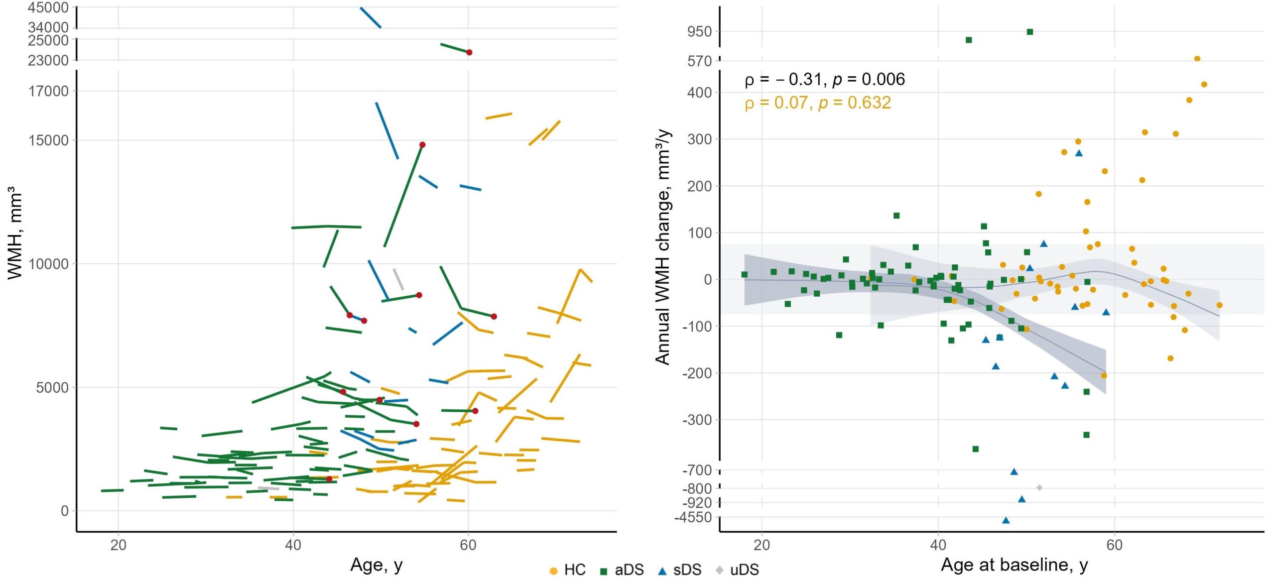
This longitudinal study included 80 adults with Down syndrome and 53 cognitively unimpaired euploid controls. WMH were segmented from serial FLAIR-MRI scans using a longitudinal-specific pipeline. We examined the effect of demographic, genetic, and clinical factors, AD clinical stages, fluid AD biomarkers, and cerebrovascular markers on annual changes in WMH volume.
Key findings
- The evolution of WMH in people with DS is variable over time and with age.
- After age 40, a growing proportion of individuals with DS showed significant WMH fluctuations, with decreases being more common than increases.
- Individuals with DS, particularly those with AD symptoms, showed a greater decline in WMH volume compared to the general population.
- Higher baseline WMH volume and the presence of microbleeds were associated with greater annual reduction of these lesions.
- This unexpected decrease in WMH, not fully explained by white matter atrophy, suggests the contribution of other mechanisms, such as neuroinflammation, to WMH dynamics in DS.
Why is this important?
People with DS present a distinct vascular risk profile compared to the general aging population; they are relatively protected from conventional vascular risk factors such as hypertension. Thus, DS offers a unique opportunity for studying cerebrovascular disease in the context of Alzheimer’s, without the confound of age-related vascular risk factors.
The findings of this study challenge the assumption that WMH in DS represent static accumulations of irreversible injury. Their dynamic and unexpected evolution points to other mechanisms, including cerebral amyloid angiopathy and inflammatory processes, that are not yet fully understood.
These insights are particularly relevant in the current landscape, in which people with DS are increasingly enrolled in clinical trials of anti-amyloid therapies. Rigorously characterizing the natural history and dynamics of cerebrovascular lesions is therefore essential to ensure the safety of these treatments and to properly guide clinical care.