
Cerebral perfusion is the amount of blood reaching a given brain region at any given time. In other words, it is the “irrigation” that supplies neurons with oxygen and nutrients.
When perfusion decreases or becomes abnormally distributed, it may indicate that an area is beginning to deteriorate, even before clinical symptoms or signs of atrophy are observed, as has been reported in Alzheimer’s disease (AD).
However, perfusion patterns in Down syndrome, a genetic form of AD, remain underexplored, as does their relationship to disease pathology. Therefore, in this study we assess perfusion using a noninvasive imaging technique to detect early AD signals and to monitor progression in people with Down syndrome.
What did we do in this study?
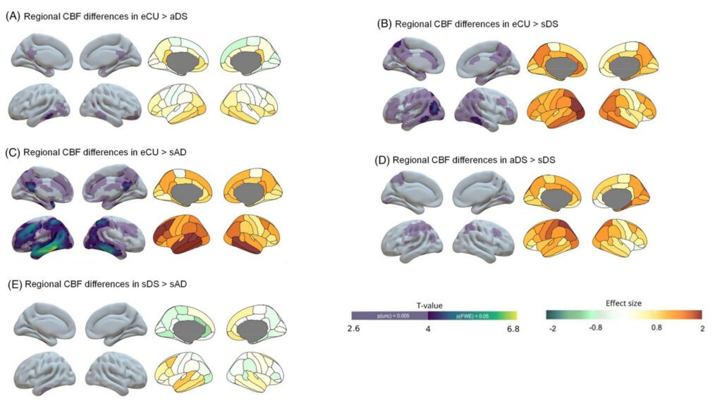
Using an MRI sequence called Arterial Spin Labeling (ASL) and processing with ASLprep, we quantified cerebral blood flow (CBF), a perfusion measure, in 51 cognitively healthy euploid controls, 54 adults with Down syndrome (34.54% symptomatic for AD), and 25 euploid patients with sporadic AD. We assessed the effects of demographic variables, clinical stage, and CSF AD biomarkers on global and regional perfusion, and we compared the topography of hypoperfusion across subgroups.
Main results
- Cerebral perfusion was reduced in Down syndrome relative to controls. Regionally, asymptomatic adults showed predominant hypoperfusion in temporoparietal areas, including the posterior cingulate cortex, inferior temporal cortex, and portions of the occipital cortex. This hypoperfusion extended into frontal regions in symptomatic stages. This pattern suggests functional progression along the AD clinical continuum.
- Within Down syndrome, individuals with AD symptoms exhibited greater parietal hypoperfusion than asymptomatic individuals.
- The hypoperfusion topography observed in symptomatic AD groups, both euploid and Down syndrome, was highly similar, reinforcing that pathophysiological mechanisms affecting cerebral blood flow are convergent between Down syndrome, associated AD and sporadic AD.
- Reductions in cerebral blood flow correlated with AD biomarkers in temporoparietal regions, underscoring the biological relevance of ASL-detected hypoperfusion as a signal of AD.
Why is this important?
Cerebral perfusion changes measured with ASL in people with Down syndrome reproduce the patterns observed in sporadic and genetic forms of Alzheimer’s disease, reinforcing their translational value. In preclinical stages, we detected early hypoperfusion in temporo-parietal regions, which extended toward frontal areas as symptoms emerged. Altogether, these results indicate that ASL, a non-invasive technique, is sensitive to detecting perfusion alterations before symptom onset in Down syndrome. Longitudinal studies are needed to establish cerebral perfusion as a progression biomarker and outcome measure in AD-targeted clinical trials in this population.