

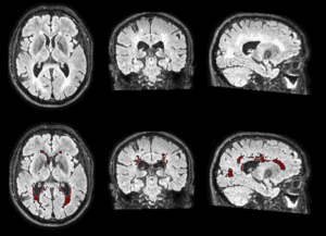
White matter hyperintensities (WMH) are one of the imaging biomarkers of small vessel disease of presumed vascular origin. These lesions can be shown as areas of increases signal on fluid-attenuated inversion recovery (FLAIR) magnetic resonance imaging. WMH are frequently found in the elderly population and in both sporadic and genetic forms of Alzheimer’s disease (AD). In Down syndrome (DS), WMH volume increases along the AD continuum. However, their spatial distribution and association with AD biomarkers remain largely unexplored.
In this study, published in the Journal of Alzheimer’s and Dementia, provides a comprehensive characterization of WMH in DS across the AD continuum.
What did we do in this study?
In this study, we segmented and extracted WMH in four concentric white matter layers and five lobar regions in 261 DS adults and 131 euploid controls. We then examined the distribution of WMH in the brain and tested associations with AD clinical stages, sociodemographic data, cerebrospinal fluid AD biomarkers, and grey matter volume.
Main results
We found that:
- WMH increased 10 years before AD symptom onset in DS.
- WMH were associated with core AD biomarkers, with the strongest association observed with NfL.
- In DS, WMH were more strongly associated with grey matter volume in parieto-temporal areas.
Relevance of this study
These findings underscore that WMH represent a core neuroimaging feature in DS and provide novel insight into the interplay between these lesions and AD processes.