The neuroimaging group, led by Dr. Alexandre Bejanin, is a multidisciplinary clinical and research team studying neurodegenerative disorders through a wide range of neuroimaging measures. The group has extensive experience with using structural magnetic resonance imaging (MRI), diffusion (DTI) and functional MRI (fMRI), but also positron emission tomography (PET) techniques to detect neurodegeneration and Alzheimer’s disease pathology. The group studies the links between these neuroimaging measurements, fluid biomarkers and cognitive decline. It is also involved in the development and implementation of new methods to detect the earliest brain changes involved in distinct neurodegenerative diseases and further our understanding of underlying pathophysiological processes. We perform research looking at distinct populations, including healthy ageing, Alzheimer’s disease in the general population but also in Down syndrome, frontotemporal lobar degeneration and dementia with Lewy Bodies.
Multiple scientific papers deriving from the work of the Neuroimaging Core have been published in high-impact journals (Aranha et al., Alzheimers Dement 2023; Alzheimers Dement 2024; Bejanin et al., JAMA Neurol 2021; Benejam et al., Alzheimers Res Ther. 2022; Illán-Gala et al., Alzheimers Res. Ther. 2022; Neurology. 2020; Montal et al., Alzheimers Dement 2021; Ann Neurol. 2021; Morcillo-Nieto et al., Alzheimmers Dement 2024; Pegueroles et al., Alzheimers Dement 2021; Zsadanyi et al., Neurology 2024 among others). This work is made possible due to various ongoing collaborations with the Nuclear Medicine Department at Hospital de Sant Pau (Dr. Albert Flotats Giralt and Dr. Valle Camacho), the Radiology Department at Hospital Clinic (Dr. Sofía González Ortiz and Dr. Nuria Bargalló), and the Radiology Department at Hospital de Sant Pau (Dr. Josep Munuera and Dr. Esther Granell).
Interested in doing your research or studies with us? Our team has several exciting active research lines, and we are constantly exploring new neuroimaging techniques to expand our understanding of changes in the brain due to neurodegenerative disease.
Structural MRI
Structural imaging can be used to detect early changes related to neurodegenerative diseases, years before the onset of symptoms. Our team utilises structural MRI to examine alterations in brain anatomy, particularly focusing on atrophy, volumetric abnormalities, and vascular lesions associated with neurodegenerative disorders. We look at whole-brain and regional atrophy, including subregions of the medial temporal lobe or the basal forebrain, structures affected by Alzheimer’s disease and other age-related pathologies. Moreover, we use structural imaging at different timepoints to study how atrophy progresses over time, and how this evolves concerning other biomarkers.
We also use structural MRI to characterise small vessel disease lesions that commonly occur in ageing and neurodegenerative diseases. Among others, we are interested in white matter hyperintensities, microbleeds, superficial siderosis, infarcts and enlarged perivascular spaces. The team implements state-of-the-art tools and pipelines, such as the Freesurfer, CAT12; Automated Segmentation of Hippocampal Subfields (ASHS) and the Lesion Segmentation Tool (LST), in our investigations of structural changes related to neurodegenerative disorders.
 Measures of cortical thickness using Freesurfer, B) Atlas of brain regions using CAT12, C) Segmentation of hippocampal subfields using ASHS, and D) Segmentation of white matter hyperintensities using LST-Lesion Prediction Algorithm (LPA).")
Diffusion MRI
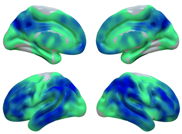
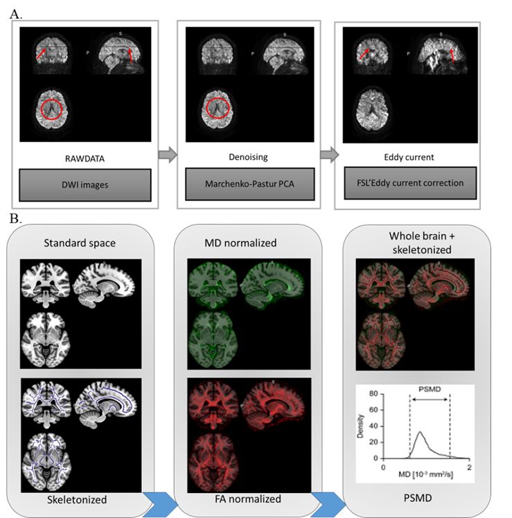
Diffusion MRI can be used to investigate alterations in brain connectivity and integrity, which can precede cognitive symptoms. Diffusion MRI techniques quantify the movement of water particles through the brain, allowing an indirect visualization of neuronal projections that connect brain regions. Diffusion MRI can also provide information on the integrity of the grey matter: our group has also developed an optimised image processing protocol to quantify the cortical mean diffusivity which reflects microstructural disorganisation and disruption of the cellular membrane and could be an early biomarker of neurodegeneration in distinct disorders. The team is also interested in using specialised diffusion measures, particularly the peak skeletonised width of mean diffusivity, to detect changes related to small vessel disease.
Functional MRI
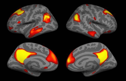
Brain network changes, measured with functional MRI, are also detectable years before symptoms appear. Functional MRI detects changes in blood flow throughout the brain and gives insight into the neural networks. By detecting fMRI alterations, we can gain crucial insights into the functional organisation of the brain throughout the process of neurodegeneration.
PET Imaging
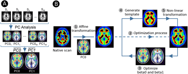
Positron emission tomography (PET) is a highly important neuroimaging technique that allows the study of metabolic changes and protein deposition that can manifest long before the appearance of first clinical symptoms of Alzheimer’s disease. PET is a non-invasive technique that allows the study of metabolic and biochemical properties of the brain. When combined with an amyloid tracer such as flutametamol or florbetapir, PET can be used to quantify the deposition of the amyloid protein in the brain. Additionally, tau-PET tracers such as Flortaucipir or PI-2620 can be used to study the presence of tau inclusions, another core pathological feature of Alzheimer’s disease. PET can also be combined with a fludeoxyglucose (FDG) tracer to study brain metabolism and determine brain areas showing abnormal glucose metabolism.
We have developed state-of-the-art methods (AMYQ) to quantify amyloid-burden across different center and without the need of an MRI or reference regions. We are currently assessing the sensitivity of FDG PET to detect early neurodegeneration in the context of distinct dementia, including in Down syndrome, and we have ongoing work to better define the pattern of tau deposition in sporadic and genetic Alzheimer’s disease.